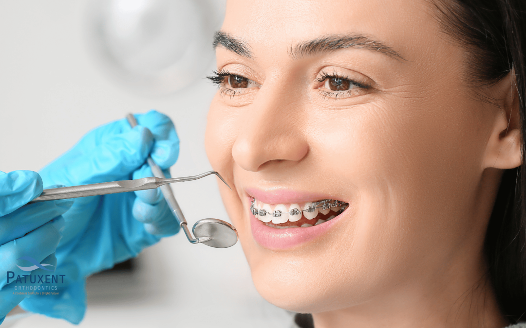
Orthodontic records consist of several diagnostic records, X-rays, photos, and films kept by your orthodontist.
They are a valuable resource for orthodontists to:
- Evaluate the entire oral cavity (including the teeth and jaws)
- Put together an effective treatment plan
- Determine facial growth patterns
Our Patuxent Orthodontics team takes great pride in keeping detailed orthodontic records for our patients. This wealth of knowledge allows us to keep constant tabs on the health of your entire oral cavity!
Let’s discover more about the various types of orthodontic records and how they benefit your oral health!
First Off, What are Orthodontic Records?
Orthodontic records are a collection of valuable documents your orthodontist assembles before starting your treatment (for example, getting braces or aligners). They assist the orthodontist in understanding your teeth and jaw health and lend an invaluable helping hand in planning your treatment.
The 8 Types of Dental Records
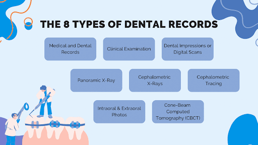
1. Medical and Dental Records
Medical and dental records contain information about the patient’s overall health, past dental work, allergies, medications, and any genetic conditions that might affect the teeth or facial structure.
2. Clinical Examination
Clinical examinations are a thorough inspection of the patient’s teeth, mouth, and jaw.
The orthodontist assesses tooth alignment, spacing, and bite function while checking for any oral health issues that need to be addressed before or during orthodontic treatment.
3. Dental Impressions or Digital Scans
Dental impressions are made by having the patient bite into a soft material that hardens into a mold. This mold is then used to create a 3D model of the teeth.
More modern practices may use a digital scanning process to create a 3D digital model.
Both methods allow the orthodontist to examine the patient’s bite and create a unique treatment plan more effectively.
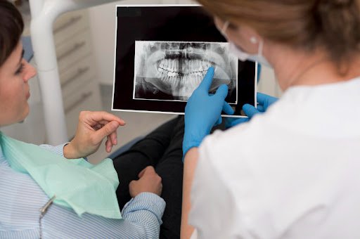
4. Panoramic X-Ray
Panoramic X-rays, also known as Panorex or simply “pano”, provide a broad view of the entire oral cavity in one image.
Unlike a traditional dental X-ray, which focuses on small, specific areas of your mouth, a panoramic X-ray shows a comprehensive view of the teeth, jaws, nasal area, sinuses, and the joints of the jaw.
This type of X-ray is particularly useful because it gives a comprehensive view of the oral cavity, allowing dentists and orthodontists to detect problems that may not be visible with a simple oral examination or an area-specific X-ray.
Issues like impacted teeth, bone abnormalities, cysts, solid growths (tumors), infections, and fractures can be easily detected using a panoramic X-ray.
5. Cephalometric X-Rays
Cephalometric X-rays, often referred to as “cephs,” are specialized radiographic examinations that capture a complete radiographic image of the side of the face.
A cephalometric X-ray differs from regular dental X-rays because they provide an image of the entire side of the head, not just the teeth. They show the relationship between the teeth, jaw, and facial profile, which makes them invaluable tools for planning and evaluating orthodontic treatment.
One of the primary uses of cephalometric X-rays in orthodontics is to assess the growth and development of the jaw in relation to the skull. This is crucial when planning orthodontic treatment, especially for young patients. The X-ray can indicate whether the patient’s jaw is aligned properly or whether an overbite or underbite is present.
6. Cephalometric Tracing
Cephalometric tracing is a method used by orthodontists to assess, evaluate, and plan treatments based on the anatomical structure of a patient’s face and jaw.
After a cephalometric X-ray is taken, a clear sheet is placed over the X-ray film. Then, your orthodontist uses a special pencil or digital tool to trace various anatomical features, including the outline of the skull, jawline, teeth, and other relevant facial structures. These points and lines create a ‘map’ of the patient’s craniofacial profile.
Cephalometric tracing is particularly important in cases that involve significant structural changes to the jaw or face, such as orthognathic surgery, severe malocclusion, or orthodontic treatment planning for children and adolescents.
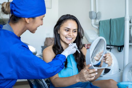
7. Intraoral & Extraoral Photos
Intraoral and extraoral photos are types of dental photographs orthodontists use to assess, diagnose, and plan treatment strategies. They are also used for documenting your treatment progress and for maintaining comprehensive records of your oral health.
Intraoral photos allow the orthodontist to capture detailed images of the teeth and the surrounding structures, like the gums, palate, and oral tissues. By using mirrors, the orthodontist can get a clear view of hard-to-see areas such as the back teeth, the inner surfaces of the upper and lower teeth and gums, and the roof of the mouth.
Intraoral photos are really helpful in identifying issues like tooth decay, gum disease, crowded teeth, or other orthodontic problems. They also provide a clear ‘before and after’ picture to demonstrate the effectiveness of your orthodontic treatment.
Extraoral photos are taken from outside the mouth. These include full-face, profile, and close-up photos of the mouth with the lips in a relaxed smiling position.
The goal of these pictures is to show the relationship between the facial appearance and the underlying skeletal and dental structures. For example, extraoral photos can reveal whether the teeth are properly aligned with the overall facial structure, whether there’s an imbalance in jaw positioning, or whether a patient’s smile is symmetrical.
8. Cone-Beam Computed Tomography (CBCT)
Cone-Beam Computed Tomography, or CBCT, represents a significant advancement in the field of dental and orthodontic imaging. It’s an X-ray machine that produces 3D images of the teeth, soft tissues, nerve pathways, and bone in a single scan.
CBCTs are particularly valuable in orthodontics because they provide a comprehensive, detailed image of the mouth, enabling precise diagnosis and treatment planning.
The CBCT machine rotates around your head, taking a series of images from different angles. These images are then processed and rendered into a single 3D image using special software. This comprehensive image can be manipulated on a computer screen, allowing the orthodontist to view the oral structures from any angle and even zoom in on specific areas of interest.
The 3 Advantages of Cone-Beam Computed Tomography Systems Over Traditional X-Rays
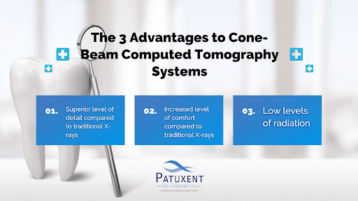
First, the level of detail provided by CBCT is far superior to traditional X-rays. It gives a complete three-dimensional view of the craniofacial structure, which is invaluable for planning complex treatments like orthognathic surgery, implant placement, or even managing impacted teeth.
Second, CBCT scans are faster and more comfortable for the patient than traditional dental X-rays. The scanning process is completed in less than a minute, and the patient doesn’t need to bite down on any plastic holders as they would with conventional X-rays.
Last, while CBCT does involve radiation, the amount is relatively low, especially when compared to a medical CT scan. This means it’s safer for patients, though it’s still primarily used in more complex cases, not for routine orthodontic check-ups.
How Do Orthodontic Records Benefit Your Treatment Plan?
Orthodontic records are the blueprint that guides the orthodontist in formulating the best course of action for achieving a patient’s dental and aesthetic goals.
Let’s dive into some of the ways these records can benefit a patient’s treatment plan!
Confirming a Diagnosis
First, orthodontic records provide a detailed picture of a patient’s oral health status. This includes not only the position and health of the teeth but also the health of the gums, the bone supporting the teeth, and the relationship of the teeth to the jaws and face.
Without this detailed picture, orthodontics are akin to flying blind — an orthodontist simply won’t have the necessary information to properly diagnose issues and plan for effective treatment.
Determining Facial Growth Patterns
Second, orthodontic records are crucial in forecasting potential future changes in a patient’s oral structure.
For example, growth prediction is an integral part of orthodontic treatment for younger patients. By using records, an orthodontist will accurately predict how a child’s jaws and teeth will develop over time and the appropriate course of treatment.

Measuring the Progress of Your Orthodontic Treatment
Last, orthodontic records help treatment providers track your progress over time.
By comparing current records to those taken earlier in the treatment process, your orthodontist will accurately measure how much your teeth have moved and if the treatment plan is on track.
This knowledge allows for the detection of any issues early on and ensures that the treatment is progressing as expected.
How Long Must an Orthodontic Office Keep Patient Records?
The length of time an orthodontic office must keep patient records can vary based on various factors, including:
- Local and state laws
- The type of record
- The age of the patient
- The guidelines set by professional orthodontic associations
It’s crucial for orthodontic offices to familiarize themselves with these regulations to ensure they are in compliance and effectively managing their records.
Local and State Laws Vary Widely
Local and state laws require that dental records be kept for a minimum of 7 to 10 years from the date of the last treatment.
This period may be extended if the patient is a minor. For instance, if the patient was a minor at the time of treatment, records are typically kept until the patient reaches the age of majority (18 years old in 48 states) plus the standard record retention period (7-10 years).
American Association of Orthodontists (AAO) Guidelines
Professional orthodontic associations often provide guidelines for their members. The American Association of Orthodontists (AAO), for example, recommends that complete patient records be retained for at least 10 years from the date the patient was last seen or until the patient turns 25.
Some Orthodontic Records are Kept Longer Than Others
Certain types of records may need to be kept longer than others. For example, records containing radiographs or study models, which can provide important diagnostic and treatment information, may need to be kept indefinitely.
Start Your Orthodontic Treatment at Patuxent Orthodontics!
Contact Patuxent Orthodontics if orthodontic care is the solution to your dental woes. Whether you want to learn more about the benefits of orthodontic care or have questions about the process, use our live chat or call (240) 802-7217 or send us a message through our contact us page to connect with our friendly staff today and book a complimentary orthodontic consultation!
Our office, located at 44220 Airport View Dr., Hollywood, MD 20636, proudly serves Maryland’s Patuxent area, as well as the Greater Washington DC area. So, if you’re residing in Hollywood, Wildewood, or Leonardtown and are looking for one of the best orthodontists in Maryland, don’t hesitate to visit our office!
We also invite you to keep up with our blog to get answers to many of the frequently asked questions about maintaining your perfect smile and follow us on Facebook and Instagram to become a part of our smiling community!
References
- “Aao Home.” American Association of Orthodontists, aaoinfo.org/. Accessed 26 June 2023.